-

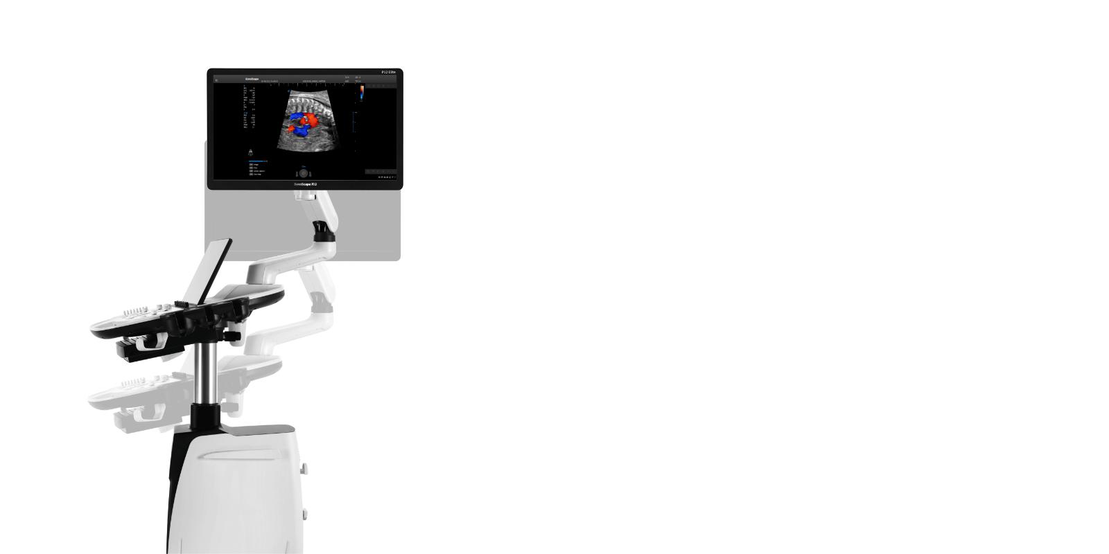
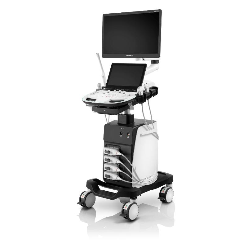
SONOSCAPE P12 Elite
Delivering Premium Solutions
-
SONOSCAPE P12 Elite
Elite Experience Meets Value
SONOSCAPE P12 Elite
SPECS
To fulfill the promise of caring for life through innovation, SonoScape offers its upgrade solution, P12 Elite, a high-definition imaging solution based on cutting-edge technologies to deliver ever more detailed images and increase your diagnostic confidence .
With comprehensive features and optimized probe configurations, the P12 Elite offers outstanding capability to handle all aspects of daily clinical practices that help build trust between clinicians and patients.
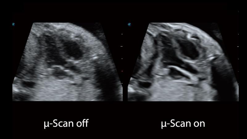
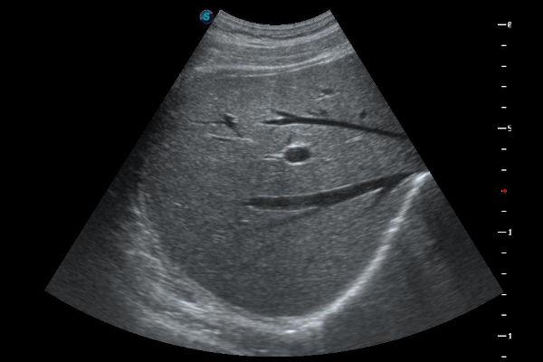
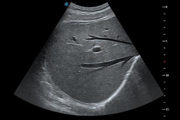
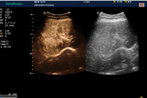
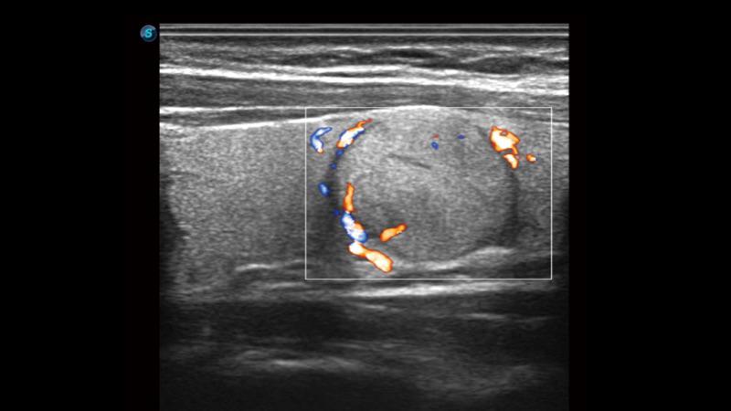
µ-Scan+
The new generation μ-Scan+ imaging technology greatly improves the visibility of organs and lesions. High definition contrast resolution will suppress speckle artifacts while maintaining real tissue architecture.
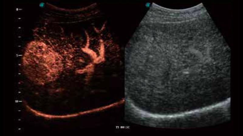
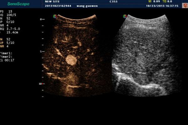
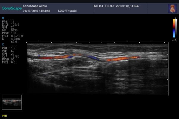
SR Flow
As an innovative new technology, SR Flow improves the ability to detect low velocity flow signals. It also improves spatial resolution and overcomes overflow to present users with real hemodymic information.
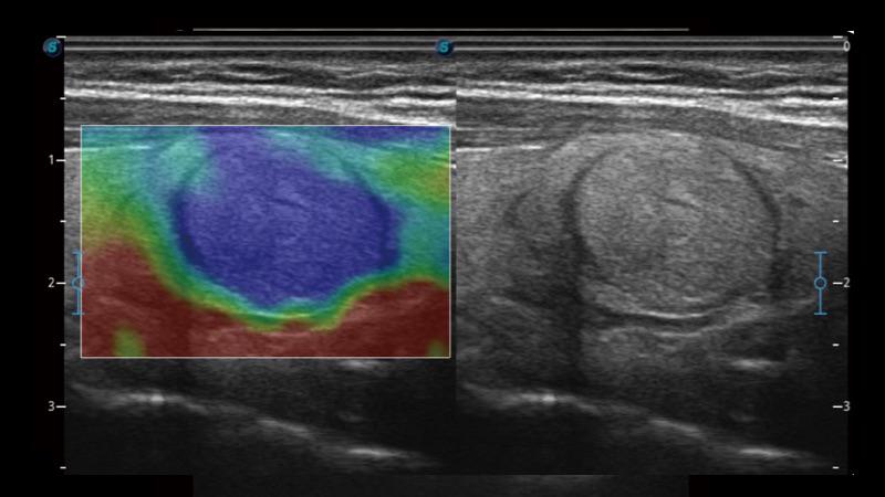
C-Xlasto
Strain elastography for evaluating tissue stiffness, professional semi-quantitative analysis with strain ratio indicating tissue elasticity
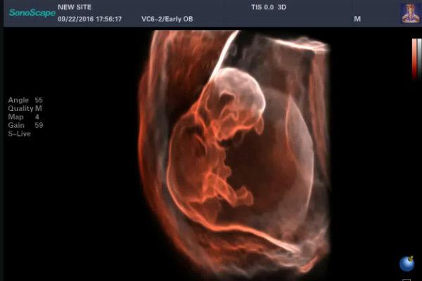
S-Live
Enhance the virtual lighting effect (especially backlight) and provide a high-quality stereoscopic image of the natural shadow and skin texture
S-Silhouette to
To see through the surface and clearly outline the contours of bones, organs, cavities, vessel walls and other internal structures.
23.8 inch LED monitor (optional), 220 degree rotation
Height-adjustable and rotatable control panel
13.3-inch tilting touch screen
Large capacity detachable battery
Multistage temperature heating gel
Sliding keyboard


SOFTWARE
-
OFF
-
ON
μ-Scan
μ-Scan uses realtime image processing algorithm to eliminate speckle and noise artifacts, enhancing tissue margins and borders by correcting discontinuity between different regions.
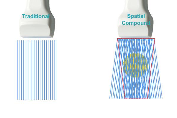
Spatial Compound Imaging
Spatial Compound Imaging utilizes several lines of sight for optimal contrast resolution, speckle reduction and border detection. Is ideal for superficial and abdominal imaging with better clarity and improved continuity of structures.
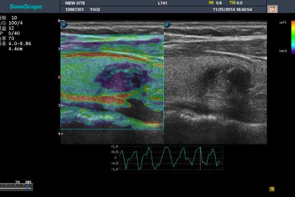
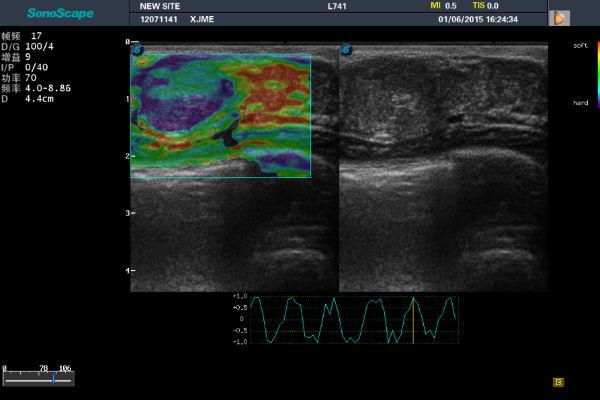
C-Xlasto
Elastography is a brand new non-invasive imaging technology. It allows the qualitative display of the elasticity of tissue according to a colorimetric scale chosen by the user. It allows to quantify the relationship between the region of interest (ROI) positioned on the suspected lesion and the surrounding tissue.
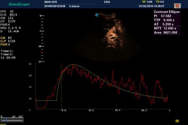
Contrast Imaging
Software for the analysis and quantification of second generation MdC.
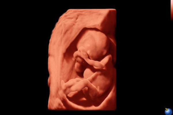
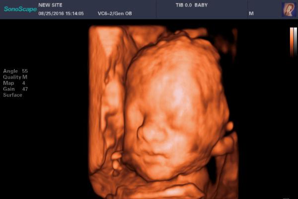
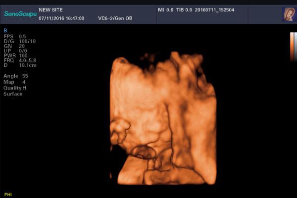
S-Live
Applies a virtual light source and advanced rendering technology that simulates the realistic skin effect.
S-Live Silouette
It allows to observe the internal anatomical structure of any volume previously acquired automatically.
Auto Face
It allows to delete the structures that interpose themselves to the profile of the fetal face.
Real Time 3D/4D
Real-time 3D imaging (4D) can intuitively show the three-dimensional structure in real time, together with the transverse and coronal planes of the various structures that we would normally not be able to acquire with 2D transducers.
Auto NT
It allows to automatically measure the nuchal translucency.
Panoramic Imaging
Real-time panoramic imaging allows visualization of larger anatomical structures than the transducer. The operator starts the sw and moves the transducer along the area of interest and the machine alligng the acquired images.
SR-Flow
• High sensitivity with directional information
• Detection of micro-vascularization and weak flows
• More realistic hemodynamic flow
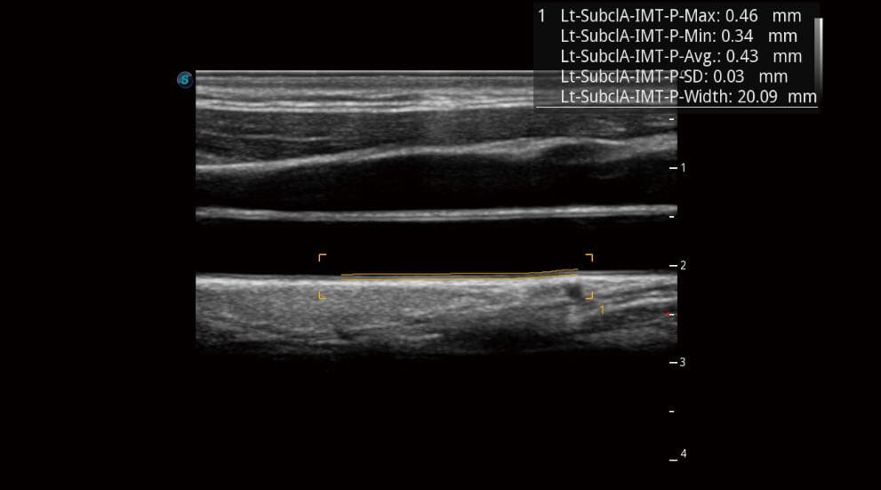
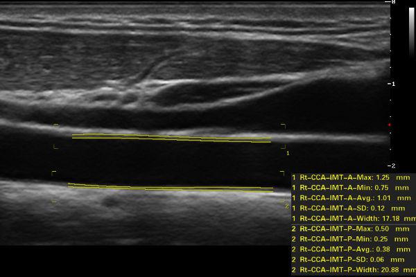
IMT
Automatically identifies the intima and measures the thickness, improving the efficiency, accuracy and repeatability of the examination.
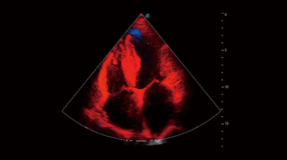
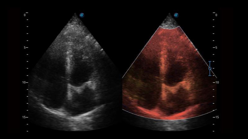
TDI
TDI can obtain information myocardial velocity, direction and time so as to analyze cardiac function more intuitively. TDI allows you to quantitatively evaluate local myocardial motion, observe myocardial velocity of different cardiac part and estimate whether there is a local lesions, as well as evaluate the early diastolic function.
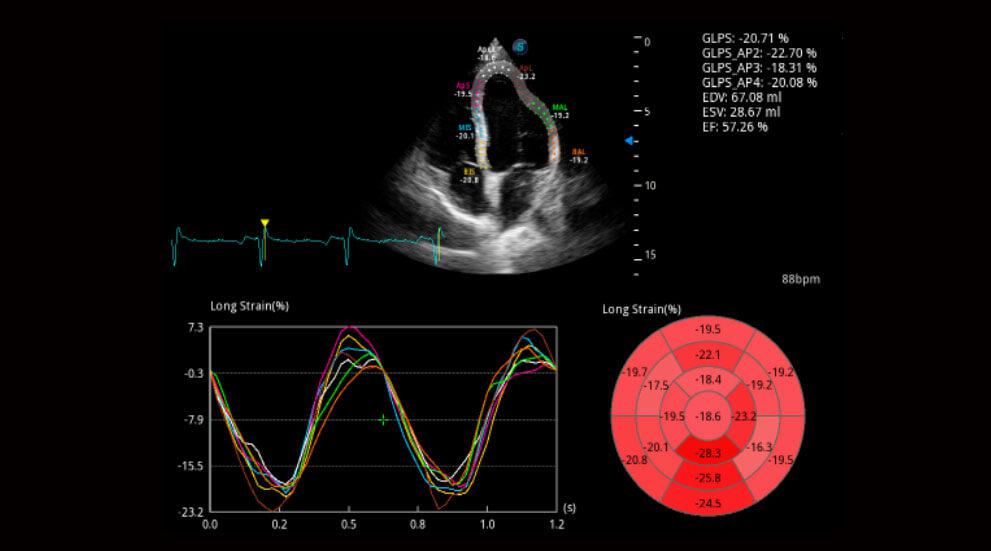
MQA
Myocardium Quantitative Analysis (MQA)
Precise quantitative measurement on myocardial mechanics is achieved by MQA based on real-time sensitive wall motion tracking. It provides global and regional assessment including strain, strain rate, displacement, velocity, etc.
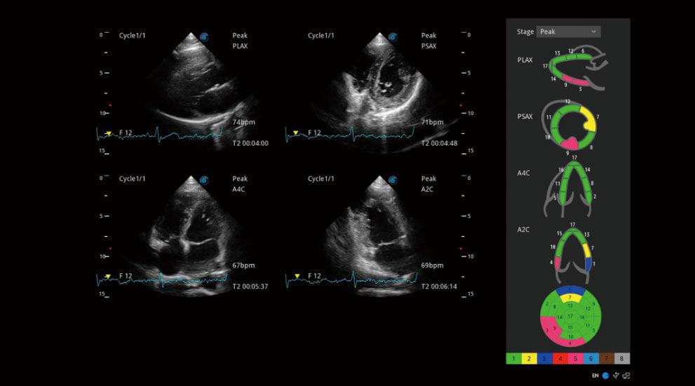
Stress Echo
Stress echo is used to diagnose coronary heart disease, evaluate coronary reserve function and myocardial ischemia, and estimate myocardial viability, providing valuable diagnostic information for PCI&CABG.
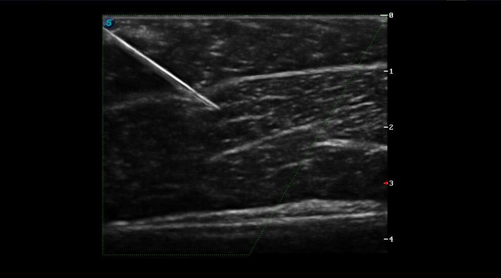
Vis-Needle
By emphasizing the visualization of the needle, it increases the safety and accuracy of biopsy procedures and other interventional procedures including nerve blocks and vascular accesses.