-
P40 Elite
Elite Experience Meets Value
-
P40 Elite
Elite Experience Meets Value
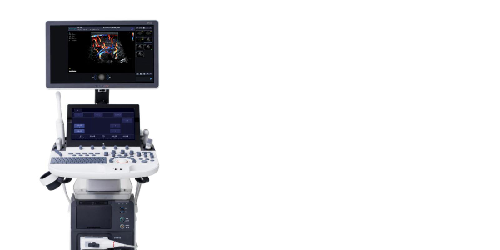
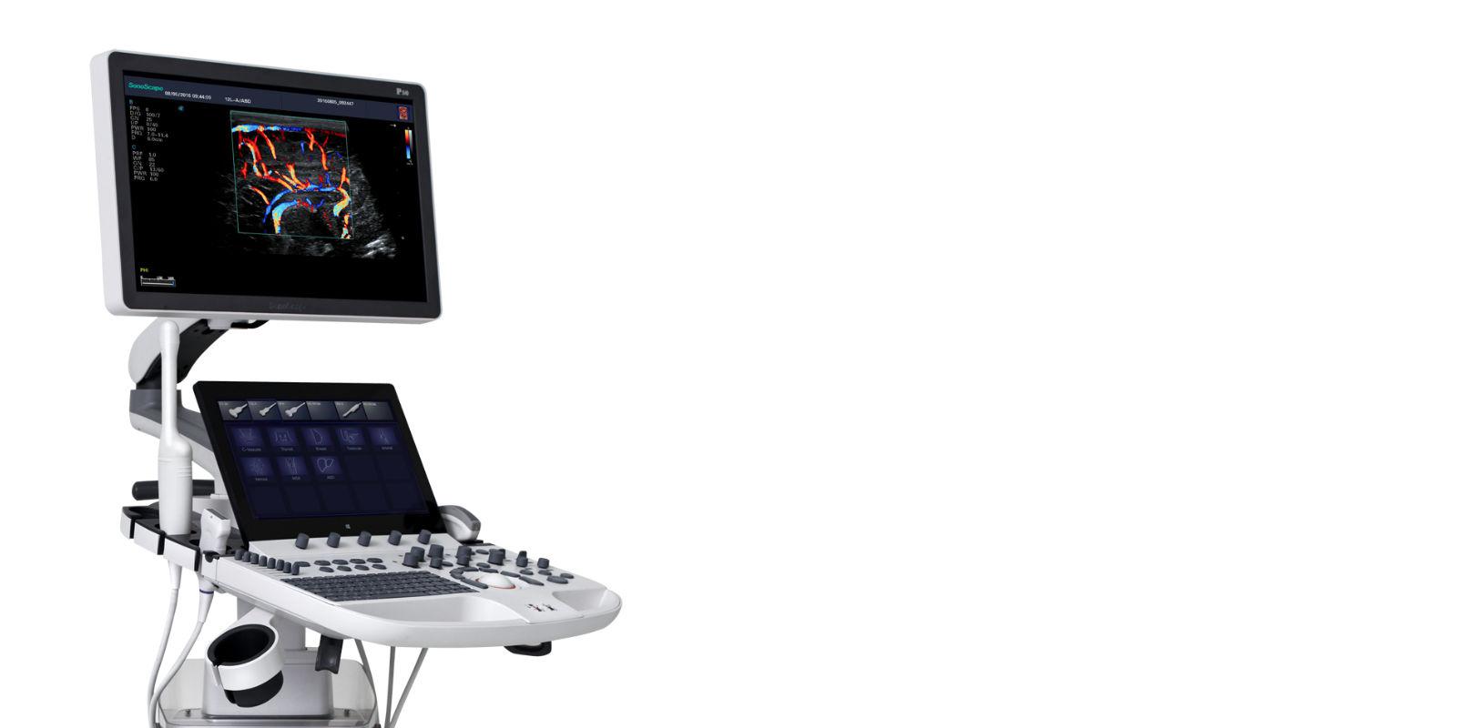
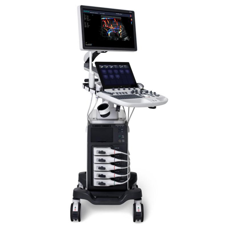
SONOSCAPE P40 Elite
SPECS
The P40 Elite system improves flexibility and effectiveness by taking it to a new level. The P40 Elite with its signal transmission and reception processors offers greater sensitivity and more accurate anatomical structure detection. In addition, the P40 Elite is equipped with a wide range of transducers that adopt innovative technologies and thus promise superior diagnostic experience.
Advanced imaging architecture that leverages the new Single Crystal probes, the system expands diagnostic capability and quality in all clinical applications.
μ-Scan
Adaptive multi-ray imaging
Dynamic color
Single crystal transducers
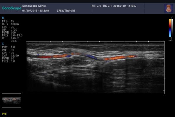
S-Thyroid is an advanced tool to detect and classify suspected thyroid lesions based on ACR TI-RADS (American College of Radiology Thyroid Imaging Reporting and Data System) guidelines. After selecting the region of interest, S-Thyroid can automatically define the boundaries of the lesion and generate a report on the characteristics of the suspected injury.
S-Brest is an advanced tool for detecting and classifying suspected breast injuries based on BI-RADS guidelines. After selecting the region of interest, S-Brest can automatically define the boundaries of the lesion and generate a report on the characteristics of the suspected injury.
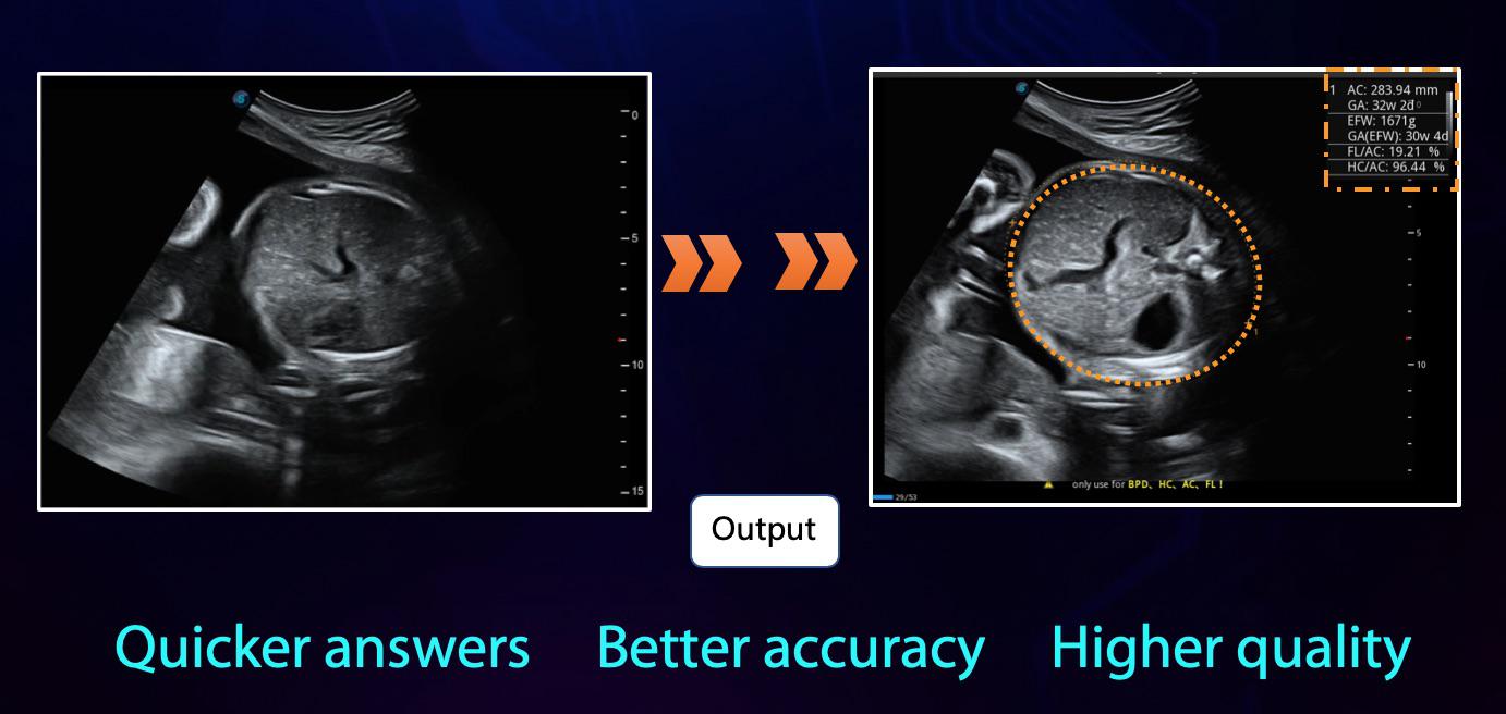
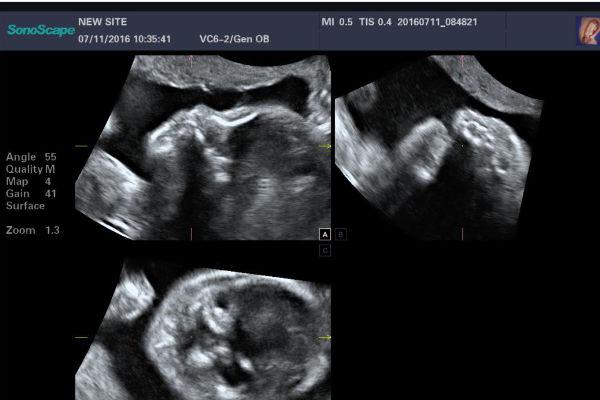
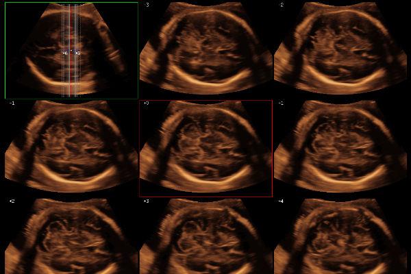
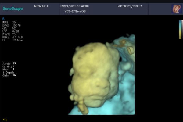
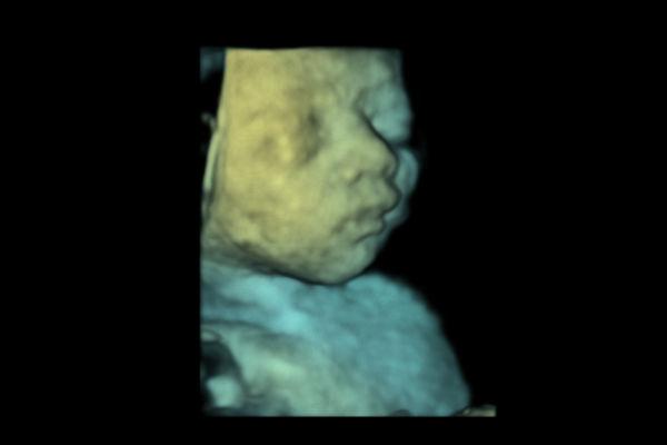
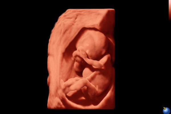
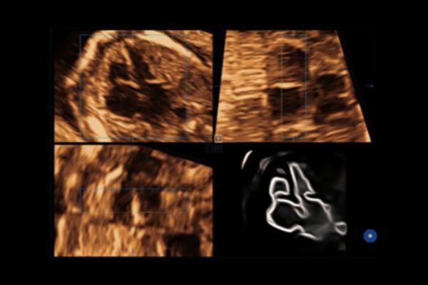
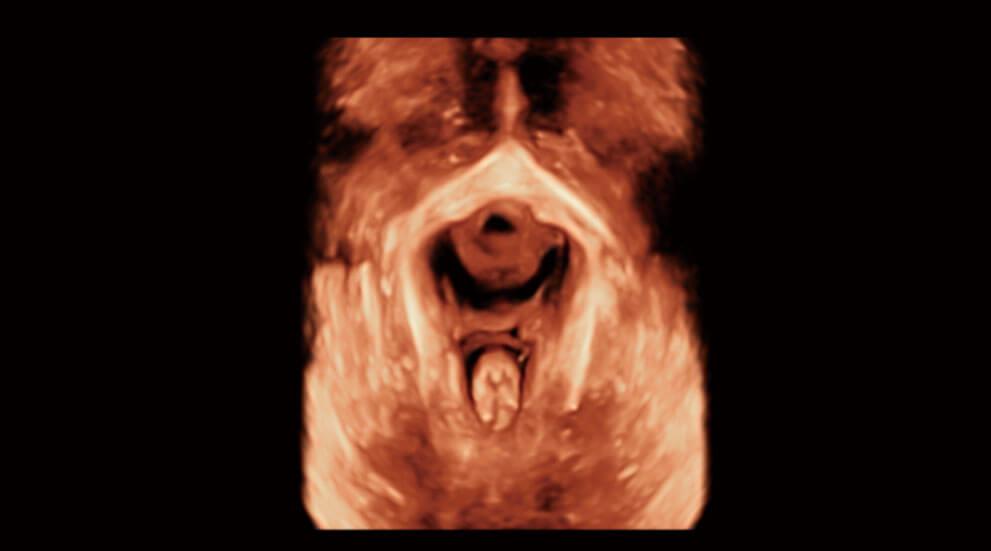
S-Fetus is a user-friendly tool that allows fully automatic and accurate detection of the most significant plans and frequently used measurements of fetal biometrics. With a fetal head cine ring, S-Fetus can extract standard planes and display measurement results in a second, greatly reducing keystrokes and required working time by several times. It is designed to turn obstetric ultrasound into a much more welcoming, faster and enjoyable experience.
S-Pelvic is an advanced tool designed to reinvent the way doctors assess pelvic floor dysfunction (PFD). Thanks to highly intelligent capabilities, full automation of pelvic floor anatomy recognition, tracking and measurement are now available and can be achieved with a single click with unprecedented ease. In addition, S-Pelvic meets the 2D automatic front compartment rating and the 3D/4D automatic lever eltus rating and takes into account both Valsalva rest and maneuvering, with the aim of covering as many steps and details as possible in the pelvic floor ultrasound and offering a complete user experience.
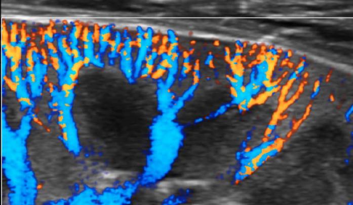
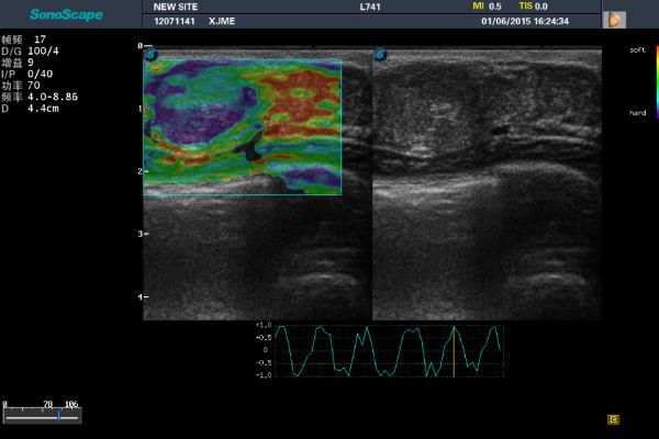
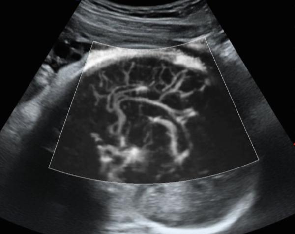
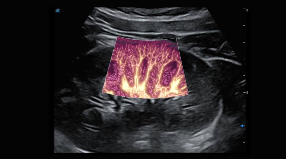
Micro F provides an innovative method for expanding the visible flow range in ultrasound, especially for the visualization of small slow-flowing vessels. By adopting an advanced adaptive filter and accumulating temporal and spatial signals, Micro F can effectively distinguish minute flow from the movement of the overlapping tissue and represent hemodynamics with increased sensitivity and spatial resolution.
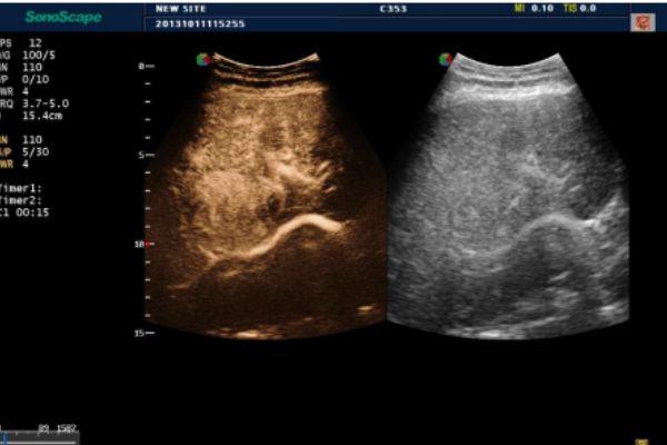
Bright Flow strengthens the definition of vessel boundaries by adding a 3D-like look to 2D color Doppler imaging. This innovative technology offers easy and improved spatial understanding and allows doctors to identify small blood flows as in a pop-off style. Luminous flux is also available for use in conjunction with other imaging modes, with the adjustable level of improvement, which offers more possibilities for clearer viewing.
Ergonomic design
21.5" LED monitor with articulated arm
13.3” adjustable touch screen
Swivel and height adjustable control panel
Integrated gel heater
Wi-Fi connection


SOFTWARE
-
OFF
-
ON
μ-Scan
μ-Scan uses realtime image processing algorithm to eliminate speckle and noise artifacts, enhancing tissue margins and borders by correcting discontinuity between different regions.
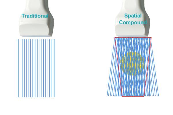
Spatial Compound Imaging
Spatial Compound Imaging utilizes several lines of sight for optimal contrast resolution, speckle reduction and border detection. Is ideal for superficial and abdominal imaging with better clarity and improved continuity of structures.
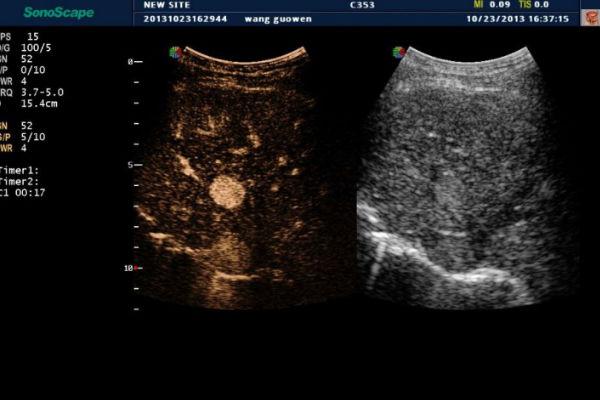
C-Xlasto
Elastography is a brand new non-invasive imaging technology. It allows the qualitative display of the elasticity of tissue according to a colorimetric scale chosen by the user. It allows to quantify the relationship between the region of interest (ROI) positioned on the suspected lesion and the surrounding tissue.
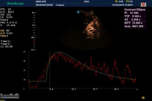
Contrast Imaging
Software for the analysis and quantification of second generation MdC.
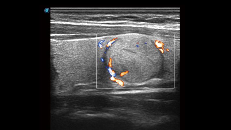
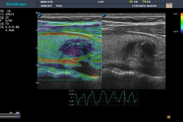
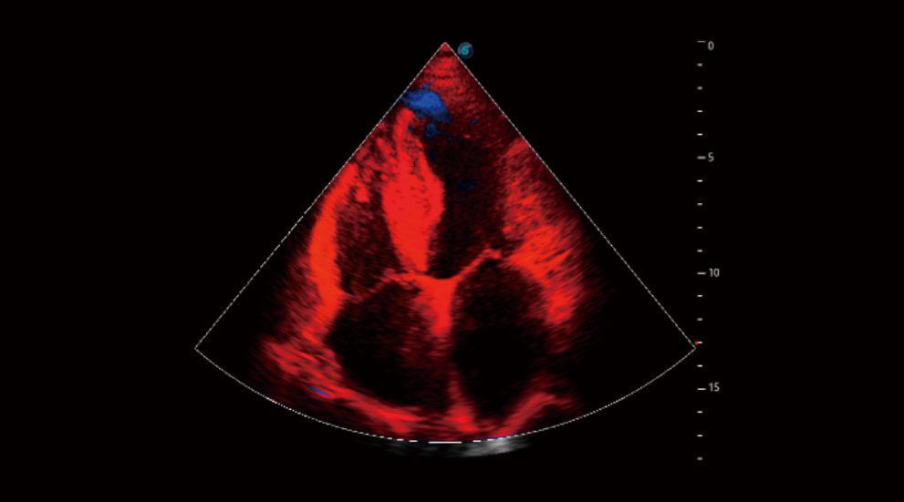
SR-Flow
• High sensitivity with directional information
• Detection of micro-vascularization and weak flows
• More realistic hemodynamic flow
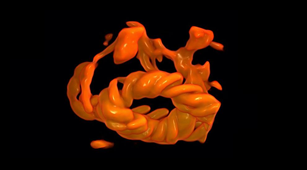
3D Color
• More intuitive and realistic flow imaging
• Improved spatial resolution of vascular networks
• Several renderings for displaying different vascular information
C-Plane
It allows to observe point-to-point the Coronal plane simultaneously with the acquisition plane and the transverse plane.
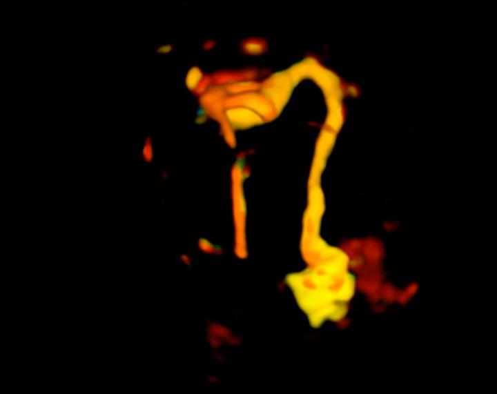
4D for Fallopian Tube
None
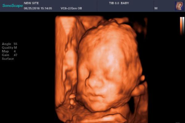
Auto Face
It allows to delete the structures that interpose themselves to the profile of the fetal face.
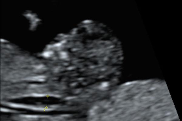
Auto NT
It allows to automatically measure the nuchal translucency.
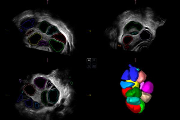
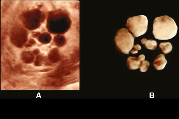
AVC-Follicle
It allows you to calculate the number and volume of follicles automatically and quickly
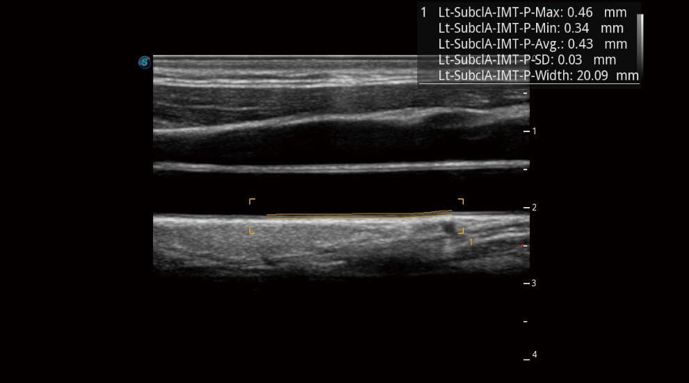
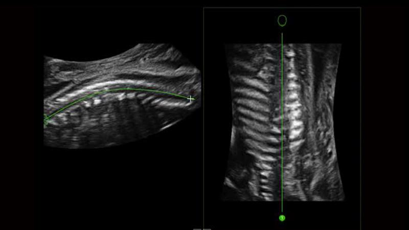
IMT
Automatically identifies the intima and measures the thickness, improving the efficiency, accuracy and repeatability of the examination.
-
None
Micro F
The adaptive Matrix E filter effectively distinguishes the blood flow of individual tissues, artifacts and flows at low speed
The algorithm helps to visualize the low blood flow in a stable and continuous way
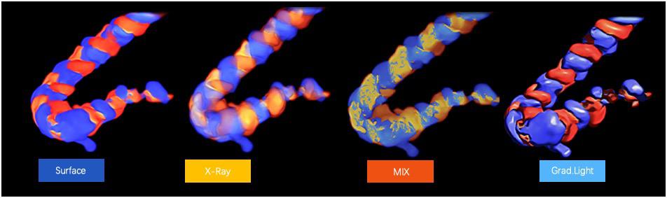
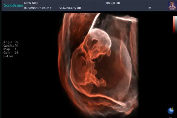
Inversion 4D
Inversion 4D provides a more in-depth evaluation of vascular and / or cystic structures by creating a three-dimensional volume of the anaechogenic structure.
Multi-Slice
Visualization of volumes in axial tomographic modality.
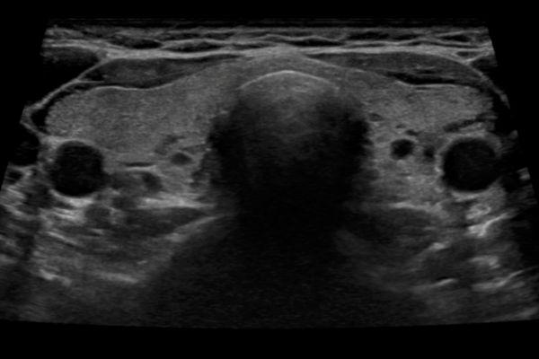
Panoramic Imaging
Real-time panoramic imaging allows visualization of larger anatomical structures than the transducer. The operator starts the sw and moves the transducer along the area of interest and the machine alligng the acquired images.
-
OFF
-
ON
Pulse Armonic Imaging
With PHI The harmonic signals are fully preserved without degradation of the acoustic information, which makes it possible to have high-level image in the visualizing of small lesions.
S-Depth
Color map show the different depth.
S-Live
Applies a virtual light source and advanced rendering technology that simulates the realistic skin effect.
S-Live Silouette
It allows to observe the internal anatomical structure of any volume previously acquired automatically.
S-Skeleton Depht
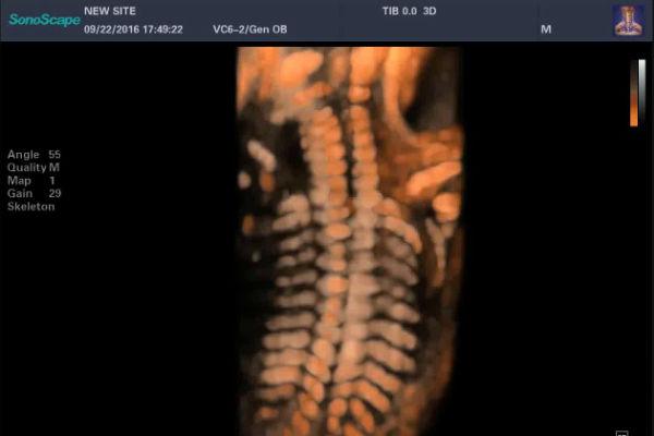
It automatically removes soft tissues to better visualize the fetal skeleton by adding a dedicated S-Depht map.
STIC
• Rapid volume acquisition in 8-12 seconds
• Beat detection for fetal anatomical structures
• Useful diagnostic tool for fetal heart disease
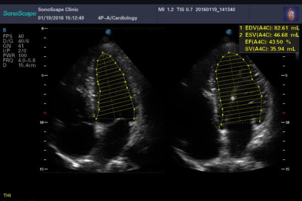
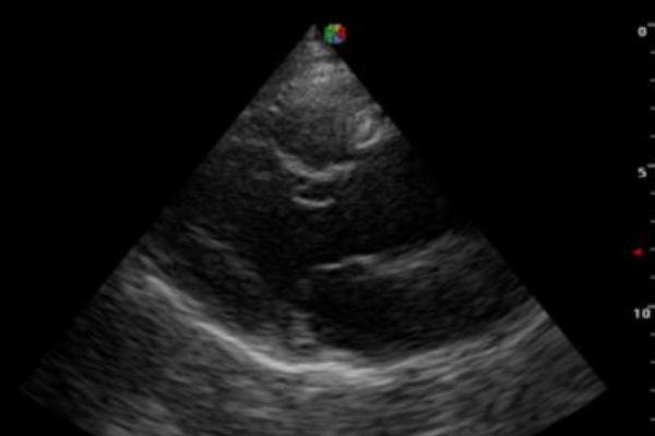
Auto EF
Automatically calculates the ejection fraction of the left ventricle through the automatic trace improving efficiency, accuracy and repeatability of cardiac examination.
Stress Echo
Stress echo is used to diagnose coronary heart disease, evaluate coronary reserve function and myocardial ischemia, and estimate myocardial viability, providing valuable diagnostic information for PCI&CABG.
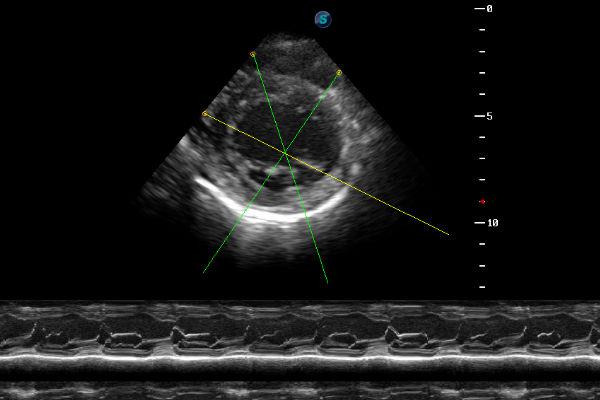
Anatomic M.Mode
Up to 3 M-Mode lines displayed simultaneously and angled according to the needs of the operator
Trapezioid Imaging
It allows to widen the field of view of linear transducers.
TDI
TDI can obtain information myocardial velocity, direction and time so as to analyze cardiac function more intuitively. TDI allows you to quantitatively evaluate local myocardial motion, observe myocardial velocity of different cardiac part and estimate whether there is a local lesions, as well as evaluate the early diastolic function.
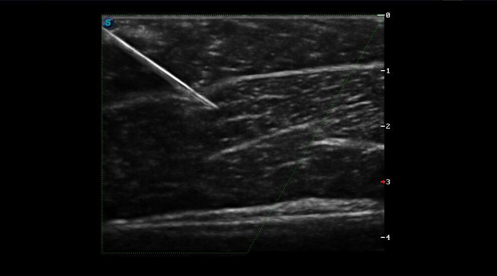
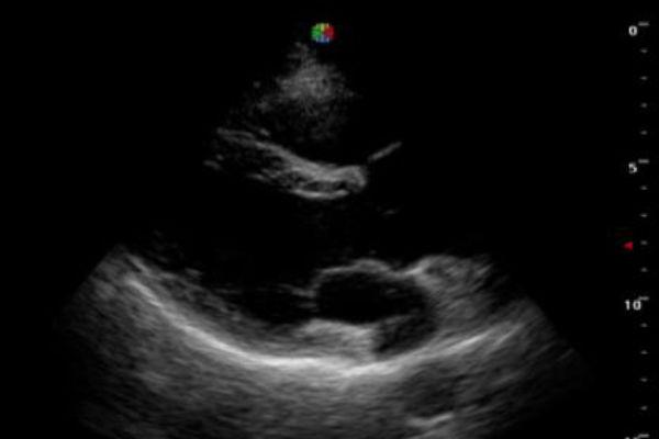
Vis-Needle
By emphasizing the visualization of the needle, it increases the safety and accuracy of biopsy procedures and other interventional procedures including nerve blocks and vascular accesses.
Bright Flow
Bright Flow
3D-like color Doppler flow strengthens boundary definition of vessel walls, without the need of using volume transducer.
Free Vue
Free Vue
Acquire any plane from 3D/4D volume data by simply defining a line in any shape. This feature makes it possible to view integral irregularly shaped structures not available in 2D imaging
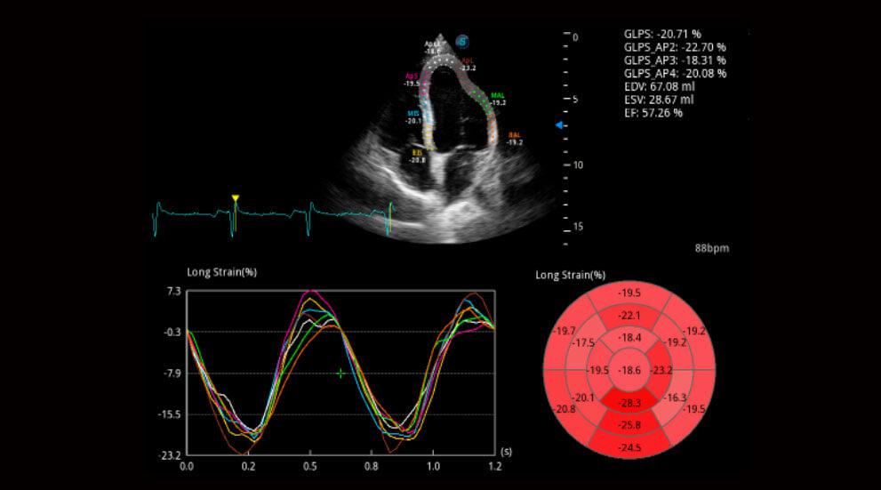
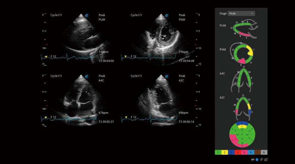
MQA
Myocardium Quantitative Analysis (MQA)
Precise quantitative measurement on myocardial mechanics is achieved by MQA based on real-time sensitive wall motion tracking. It provides global and regional assessment including strain, strain rate, displacement, velocity, etc.
Pelvic Floor
Pelvic Floor Imaging
Working in conjunction with specialized transvaginal probes, both 2D and volume imaging are available with thorough evaluation in viewing pelvic anatomy like muscles, bladder, uterus, etc.