-
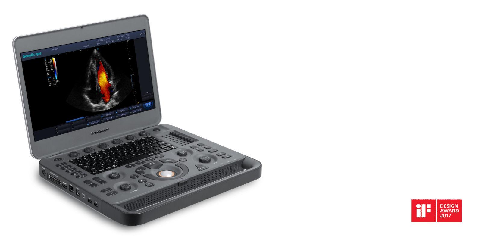
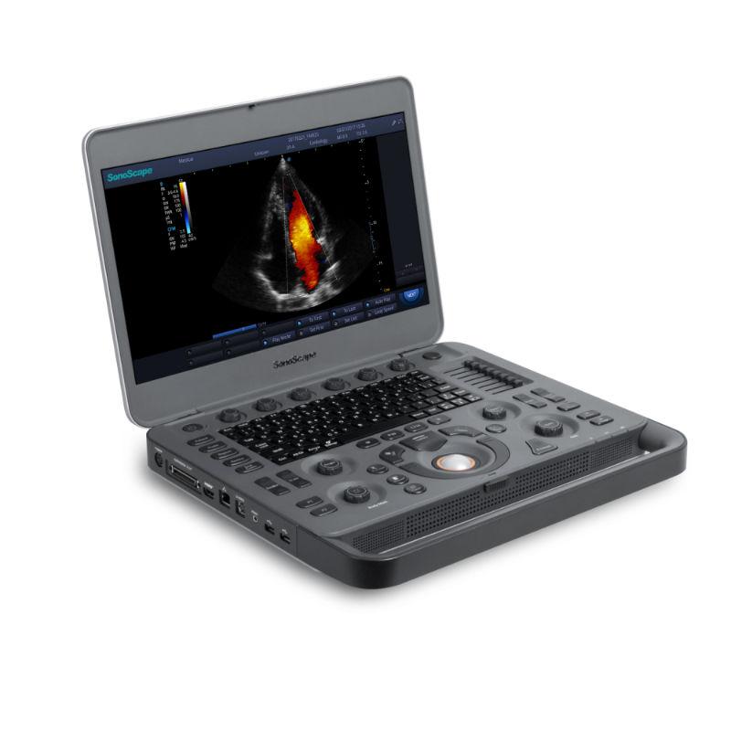
SONOSCAPE X5
Delivering Premium Solutions
-
SONOSCAPE X5
Delivering Premium Solutions
SONOSCAPE X5
SPECS
Imaging Enhancement
The new platform X5 provides, due to its improved imaging capabilities, an accurate diagnosis for an overall increased workflow.
- Multi-beam processing technology
- Spatial compound imaging
- μ-Scan image processing technology
- Pulse inversion harmonic imaging
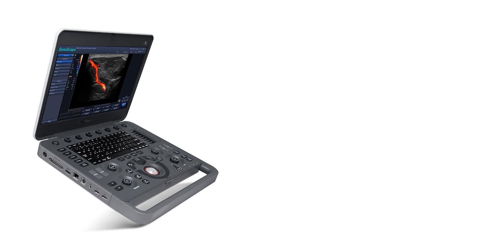
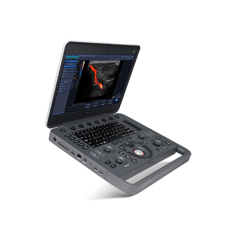
Mobility
- 15.6 inch wide range folding angle and wide viewing angle high definition monitor
- Extended connector to 3 probes
- Quick boot up time
- Built-in battery support 3 hours continuous scanning
- Integrated trolley with height adjustment
- Wi-Fi and Bluetooth wireless connection
User Friendly
- One Button automatic optimization
- Large image display region
- Anti-reflective screen and auto brightness adjustment
- User-defined Operations
Extensive Applications
- Crystal clear imaging quality makes X5 ideal for various applications.


SOFTWARE
-
OFF
-
ON
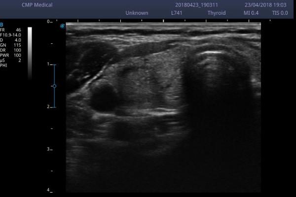
μ-Scan
μ-Scan uses realtime image processing algorithm to eliminate speckle and noise artifacts, enhancing tissue margins and borders by correcting discontinuity between different regions.
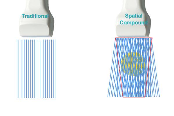
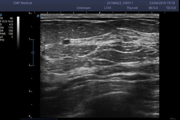
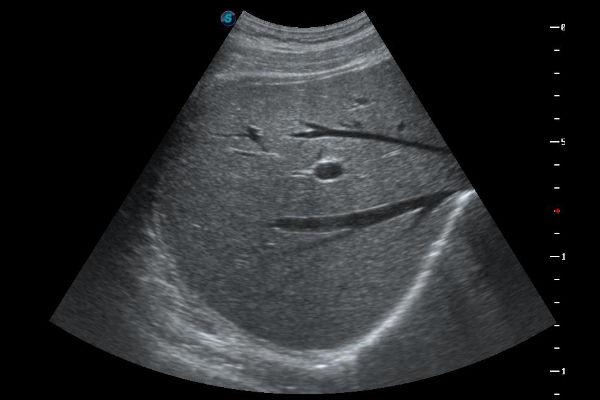
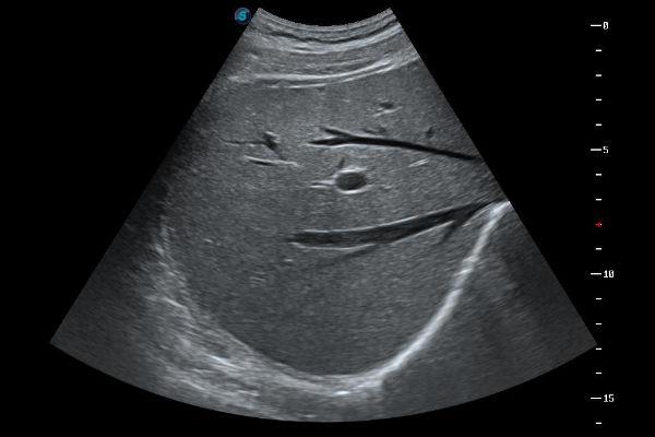
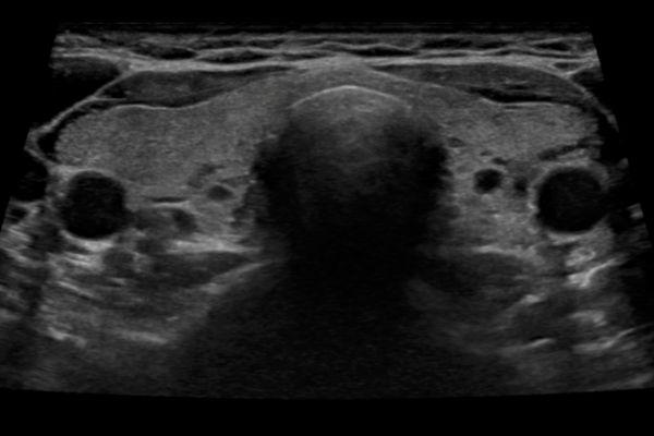
Spatial Compound Imaging
Spatial Compound Imaging utilizes several lines of sight for optimal contrast resolution, speckle reduction and border detection. Is ideal for superficial and abdominal imaging with better clarity and improved continuity of structures.
-
OFF
-
ON
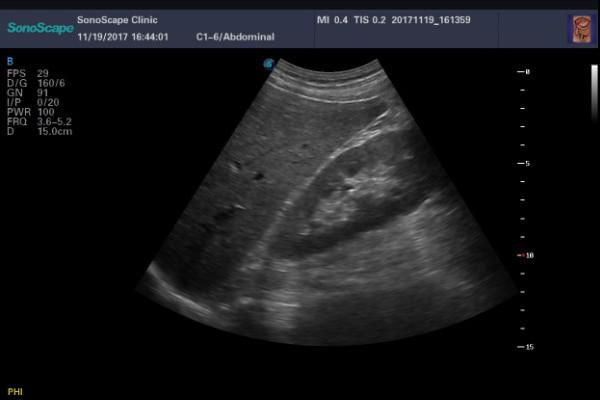
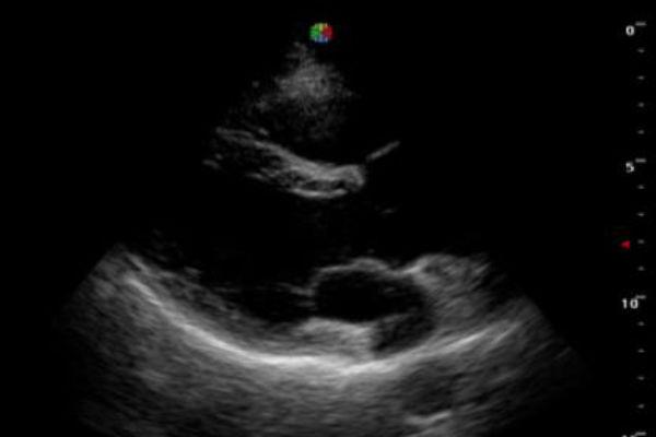
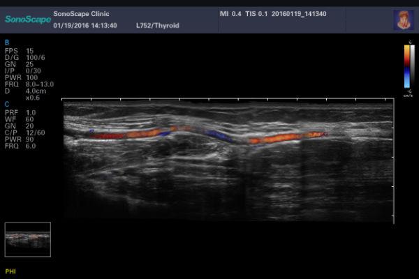
Pulse Armonic Imaging
With PHI The harmonic signals are fully preserved without degradation of the acoustic information, which makes it possible to have high-level image in the visualizing of small lesions.
Trapezioid Imaging
It allows to widen the field of view of linear transducers.
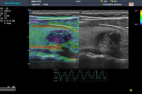
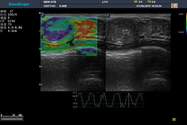
C-Xlasto
Elastography is a brand new non-invasive imaging technology. It allows the qualitative display of the elasticity of tissue according to a colorimetric scale chosen by the user. It allows to quantify the relationship between the region of interest (ROI) positioned on the suspected lesion and the surrounding tissue.
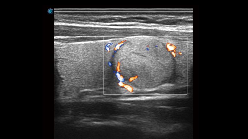
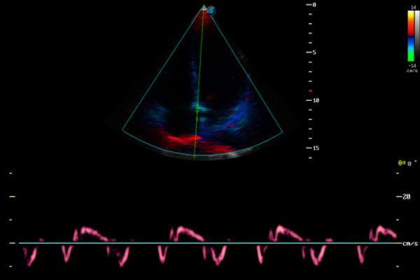
SR-Flow
• High sensitivity with directional information
• Detection of micro-vascularization and weak flows
• More realistic hemodynamic flow
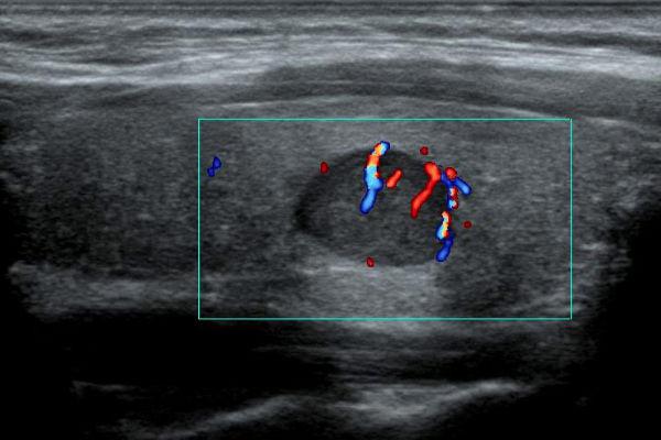
Dynamic Color
HD-Flow. With Dynamic Color the sonographers can easily see in detail very small veins and slower velocities for detailed blood flow.
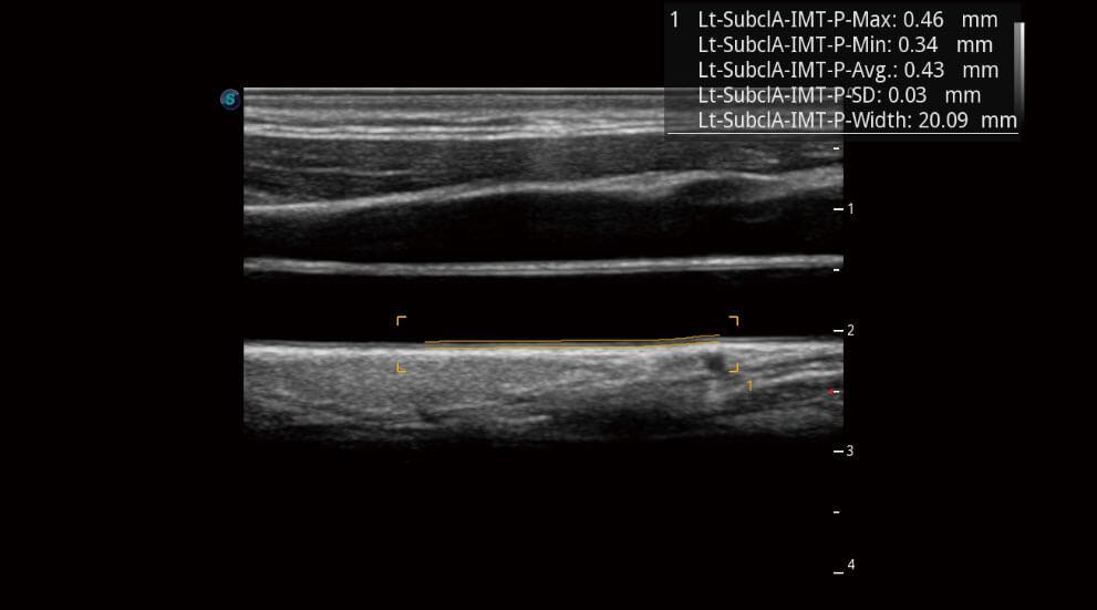
IMT
Automatically identifies the intima and measures the thickness, improving the efficiency, accuracy and repeatability of the examination.
Panoramic Imaging
Real-time panoramic imaging allows visualization of larger anatomical structures than the transducer. The operator starts the sw and moves the transducer along the area of interest and the machine alligng the acquired images.
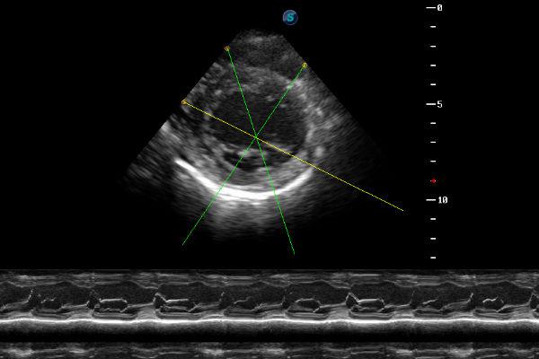
Anatomic M.Mode
Up to 3 M-Mode lines displayed simultaneously and angled according to the needs of the operator
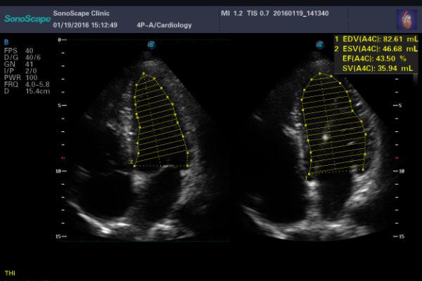
Auto EF
Automatically calculates the ejection fraction of the left ventricle through the automatic trace improving efficiency, accuracy and repeatability of cardiac examination.
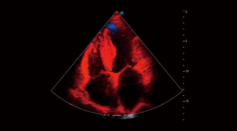
TDI
TDI can obtain information myocardial velocity, direction and time so as to analyze cardiac function more intuitively. TDI allows you to quantitatively evaluate local myocardial motion, observe myocardial velocity of different cardiac part and estimate whether there is a local lesions, as well as evaluate the early diastolic function.
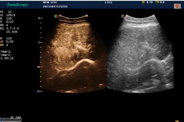
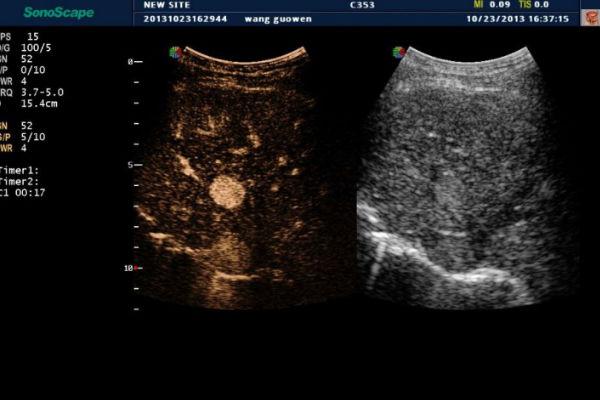
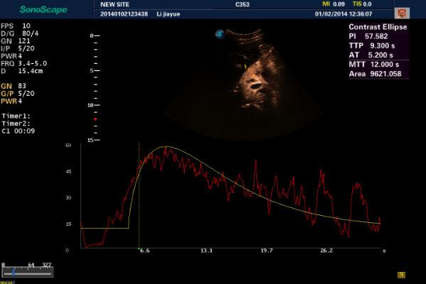
Contrast Imaging
Software for the analysis and quantification of second generation MdC.
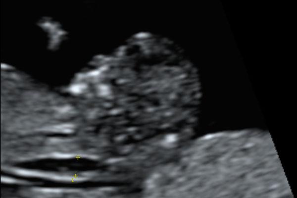
Auto NT
It allows to automatically measure the nuchal translucency.
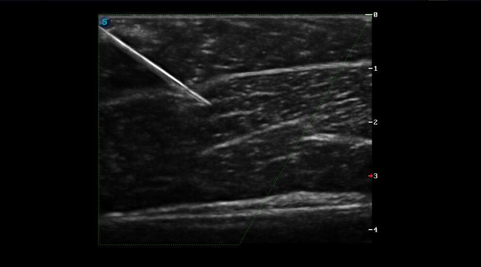
Vis-Needle
By emphasizing the visualization of the needle, it increases the safety and accuracy of biopsy procedures and other interventional procedures including nerve blocks and vascular accesses.